Title
X-Ray Diagnosis and Treatment by Bythell & Barclay (1912)
Author
W. J. S. Bythell and A. E. Barclay
Image

Description
This remarkable medical book, X-Ray Diagnosis and Treatment, was published in 1912 as part of the Oxford Medical Publications series. Written by physicians W. J. S. Bythell and A. E. Barclay, it offered an authoritative guide to the emerging field of radiology—a field still in its infancy just 17 years after Wilhelm Röntgen’s discovery of X-rays.
The book covers diagnostic and therapeutic uses of X-rays, providing insight into early 20th-century medical thought and practice. From detailed anatomical images to diagnostic methods for bone injuries, this volume captures the transitional moment when medicine was moving from physical examination to visual, image-based diagnosis.
Condition
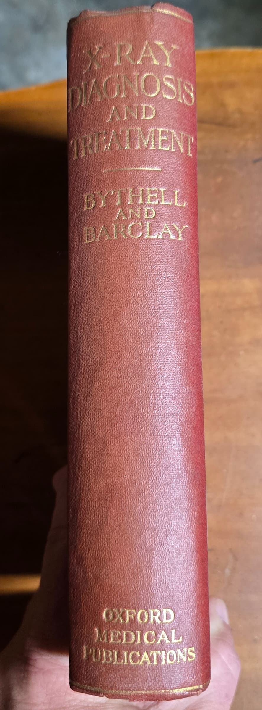
This book is in good vintage condition, with a sturdy red hardcover binding and gilt lettering on the spine. Some slight fading and age-related wear are visible on the cover, but the binding remains tight. The black-and-white X-ray images inside are crisp and haunting, adding to the historical intrigue.
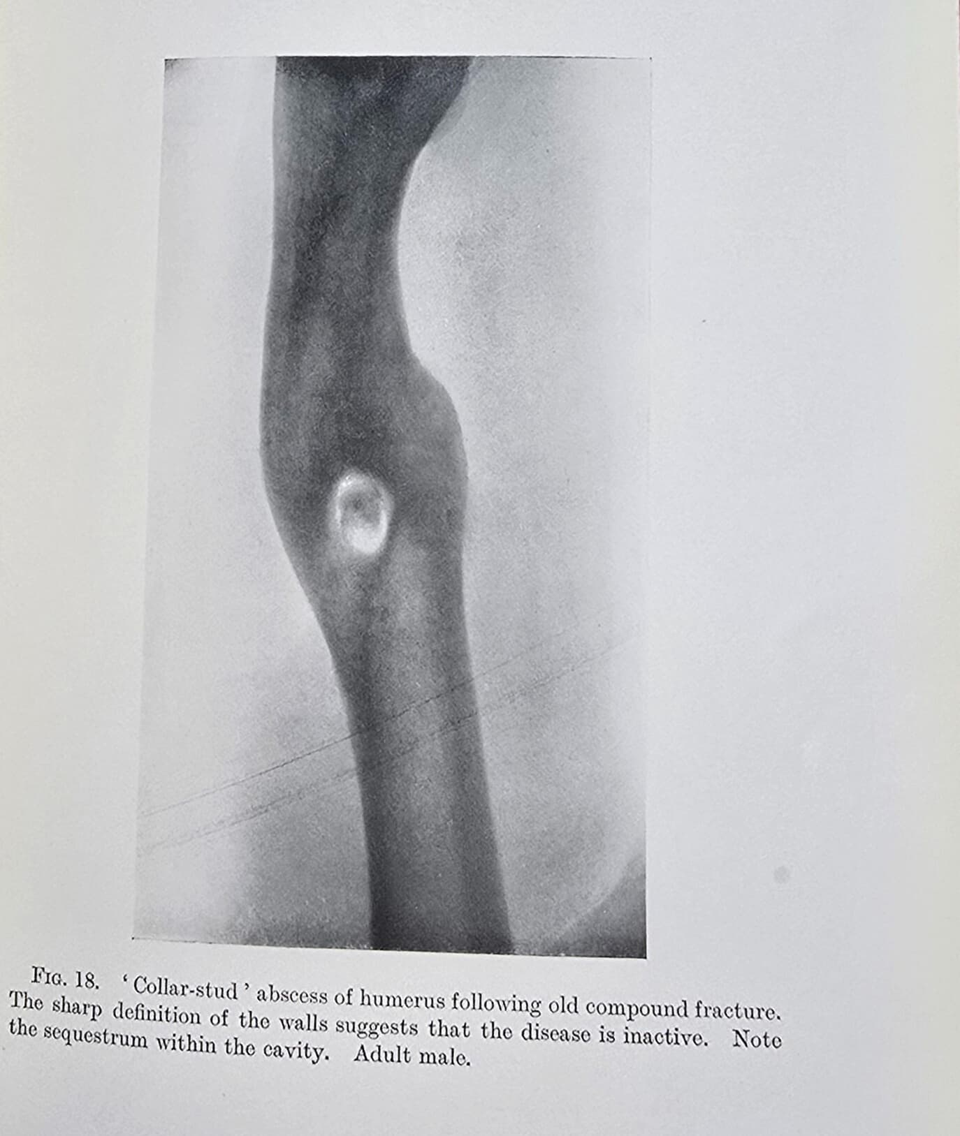
Gallery - Includes images of the striking red cover, detailed close-ups of the title and spine, and interior photographs showcasing early X-ray imagery—like the “collar-stud” abscess of the humerus, a testament to the power of these new diagnostic tools.
Historical context
Bythell and Barclay’s work sits at the dawn of radiology, when X-rays were seen as both miracle and mystery. In an era of medical experimentation, this book was a lifeline for general practitioners navigating the uncertain terrain of early 20th-century medicine. It illustrates how rapidly X-ray technology was adopted, and how it forever changed the diagnostic landscape.
Curious Facts, Ephemera, and Trivia
X-rays were so novel in the early 1900s that some patients and doctors feared they might cause invisible damage to the body—ironic, given what we know now about radiation safety!
This edition was printed by Henry Frowde at the Oxford University Press and Hodder & Stoughton, London.
The images inside reflect the earliest use of X-rays for therapeutic as well as diagnostic purposes—a practice that laid the groundwork for modern radiotherapy.
Excerpt
“Fig. 18. ‘Collar-stud’ abscess of humerus following old compound fracture. The sharp definition of the walls suggests that the disease is inactive. Note the sequestrum within the cavity. Adult male.”
—From the book’s photographic plate section
Why it is in the Cabinet
This antique medical textbook is a window into the moment when physicians began peering inside the human body with unprecedented clarity. It represents not just scientific progress, but also the hope and uncertainty that always accompany new discoveries. It’s a proud part of the Cabinet of Curiosities, reminding us how far we’ve come—and how much we owe to the pioneers who first harnessed the power of the invisible.
See also: Diseases of the Nose and Throat | Antique Wooden Crutch
Support Dr. Bebout’s Cabinet of Medical Curiosities
If you enjoy the history, the oddities, and the effort, help keep this cabinet open. Every little bit helps preserve and share the strange wonders of medicine's past.
Buy Me a Ko-fi ☕ Buy Me a Coffee ☕ Tip via PayPal 💵