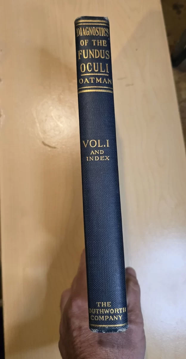
Title
Diagnostics of the Fundus Oculi (1920, 3-Volume Set)
Author
Edward L. Oatman, M.D.
Surgeon, Manhattan Eye, Ear and Throat Hospital; Brooklyn Eye and Ear Hospital; Consulting Ophthalmic Surgeon, Nyack Hospital and St. Mary’s Hospital, Waterbury, Connecticut.
Image
Description
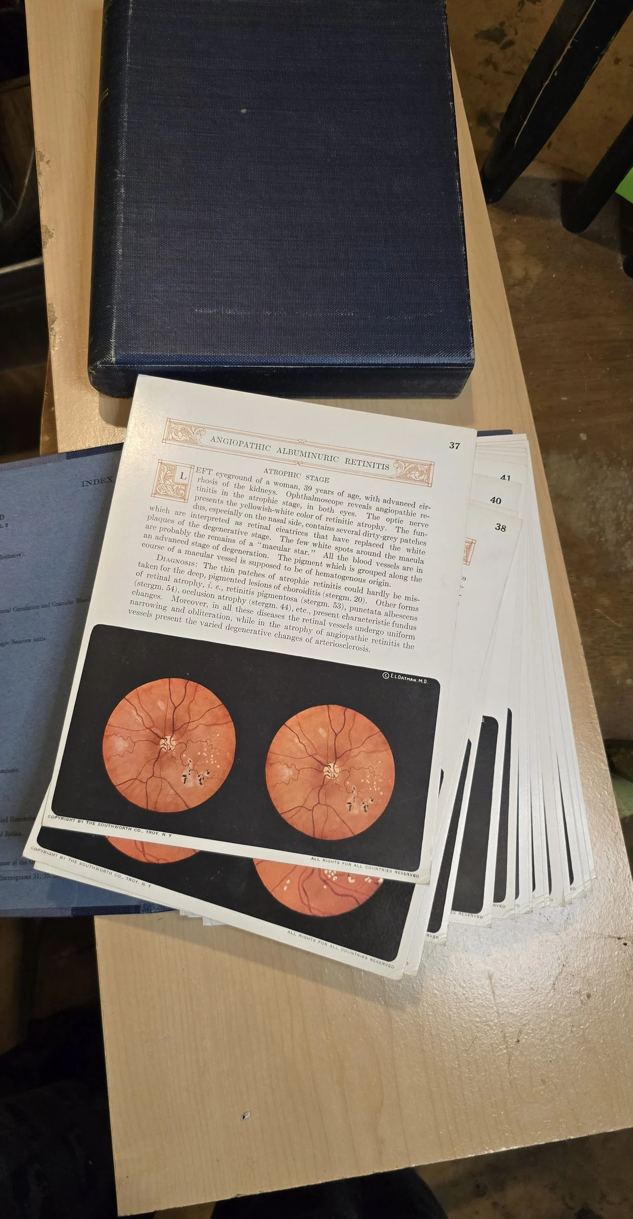
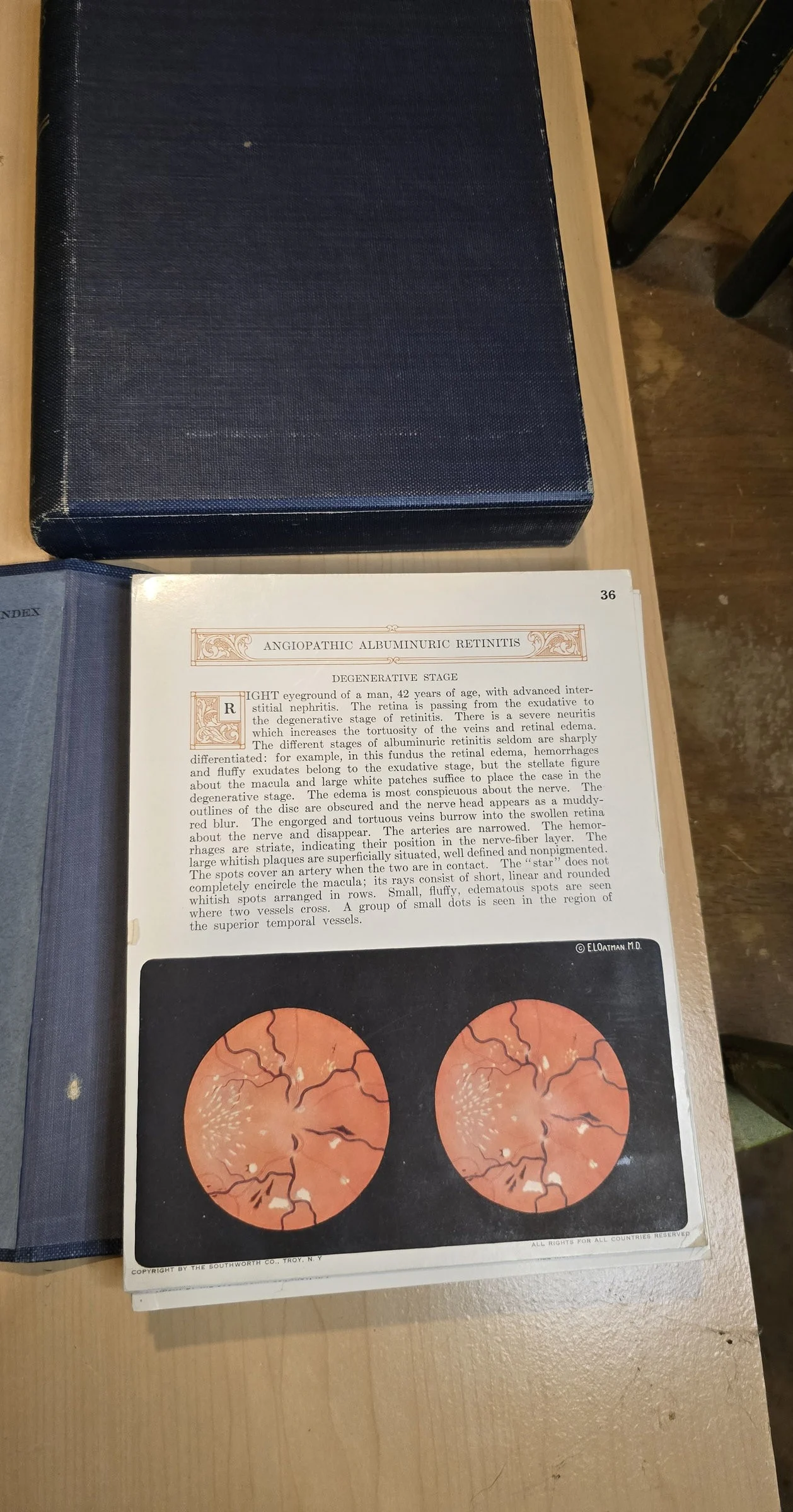
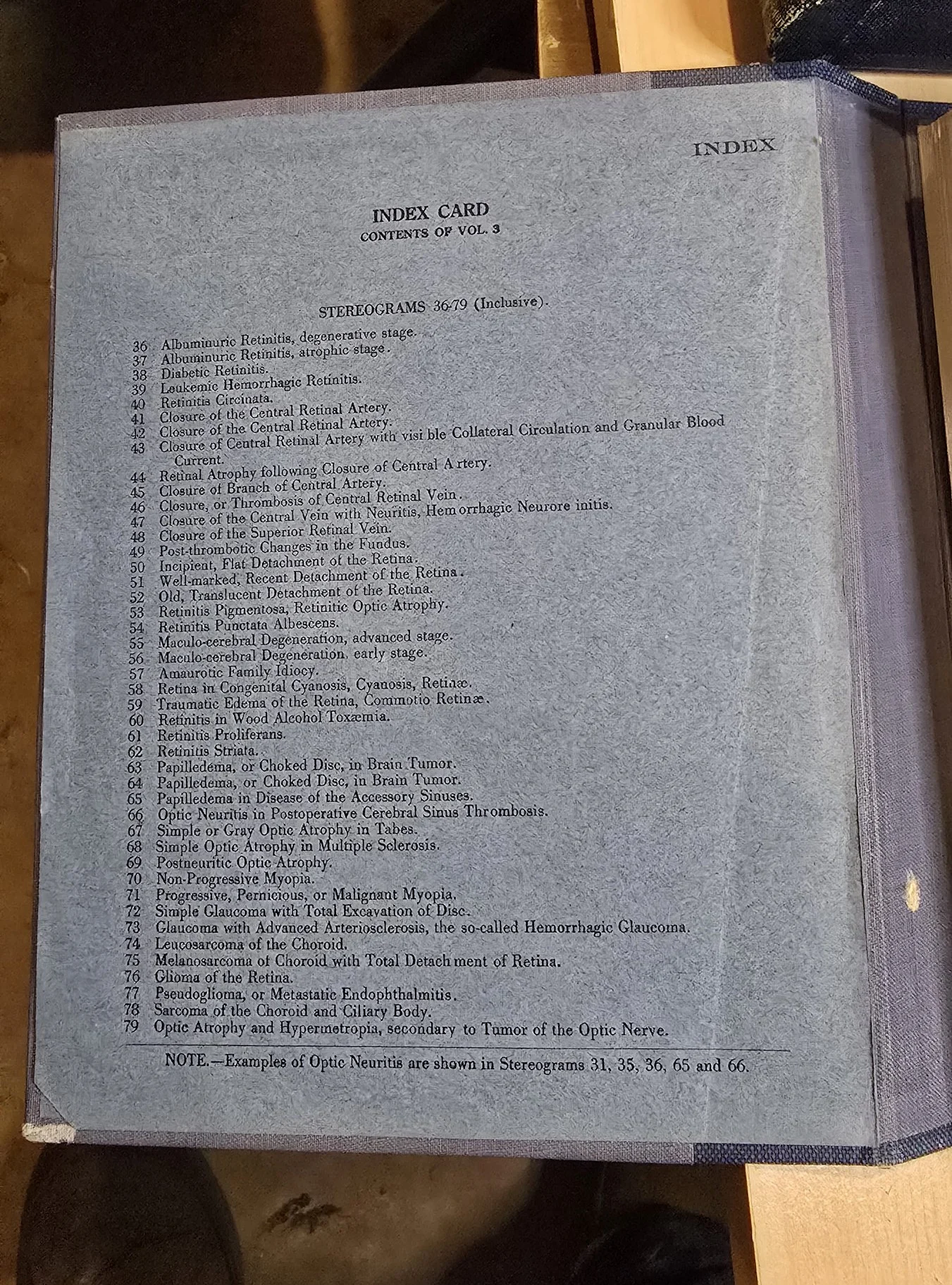
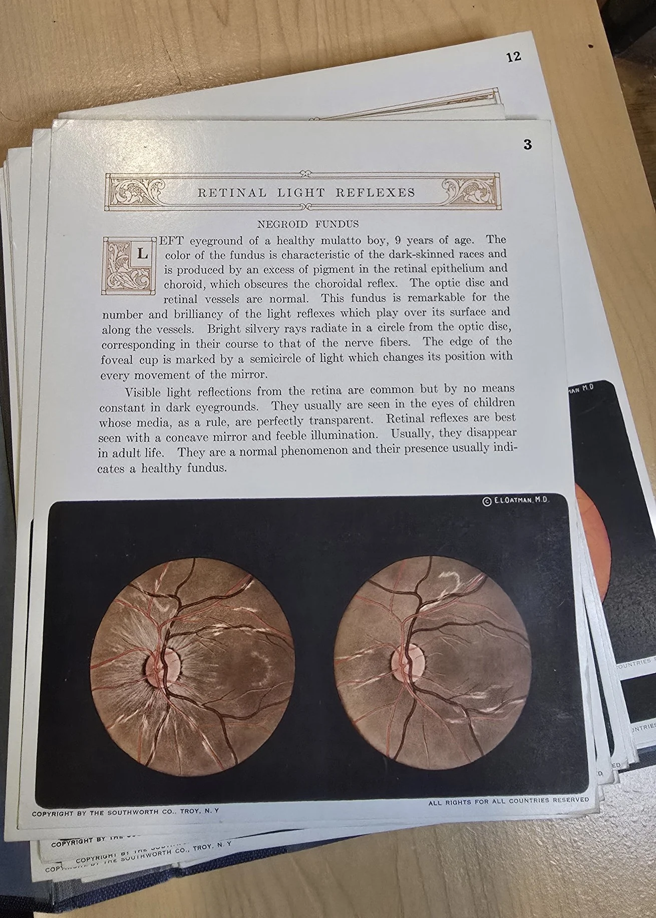
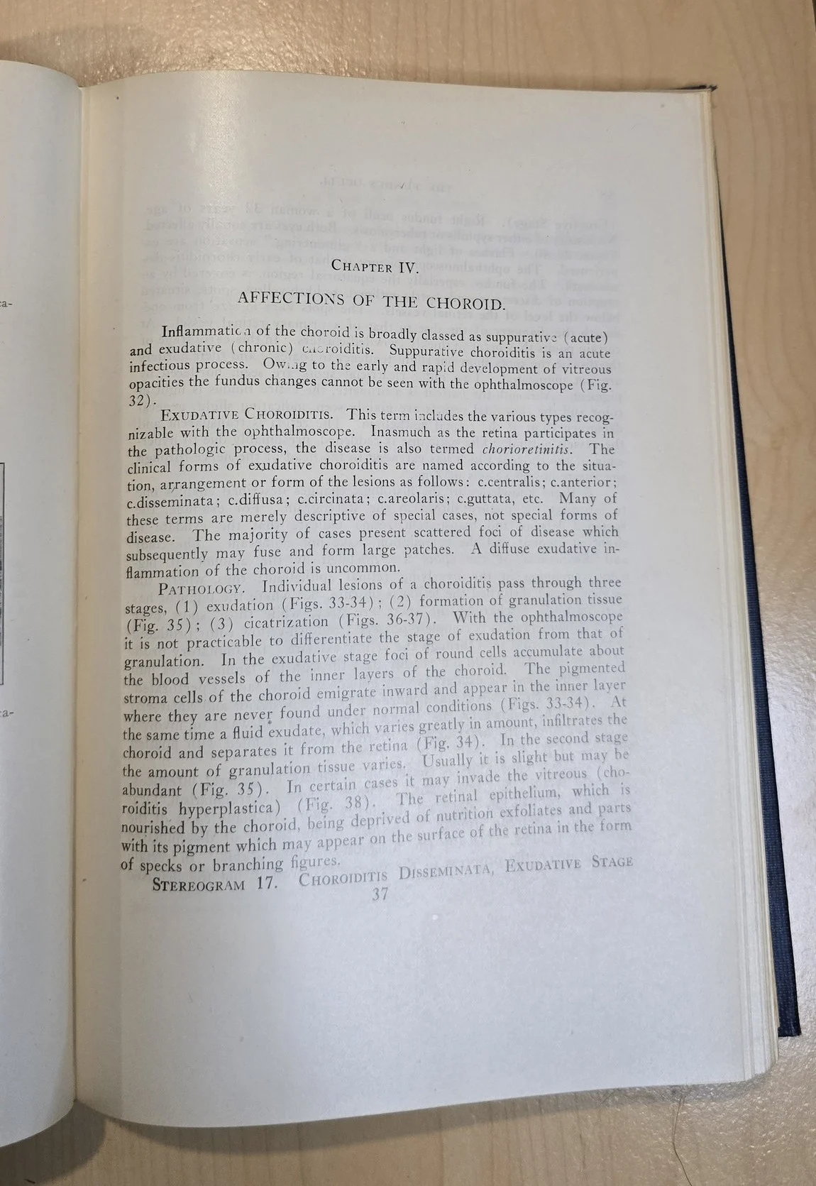
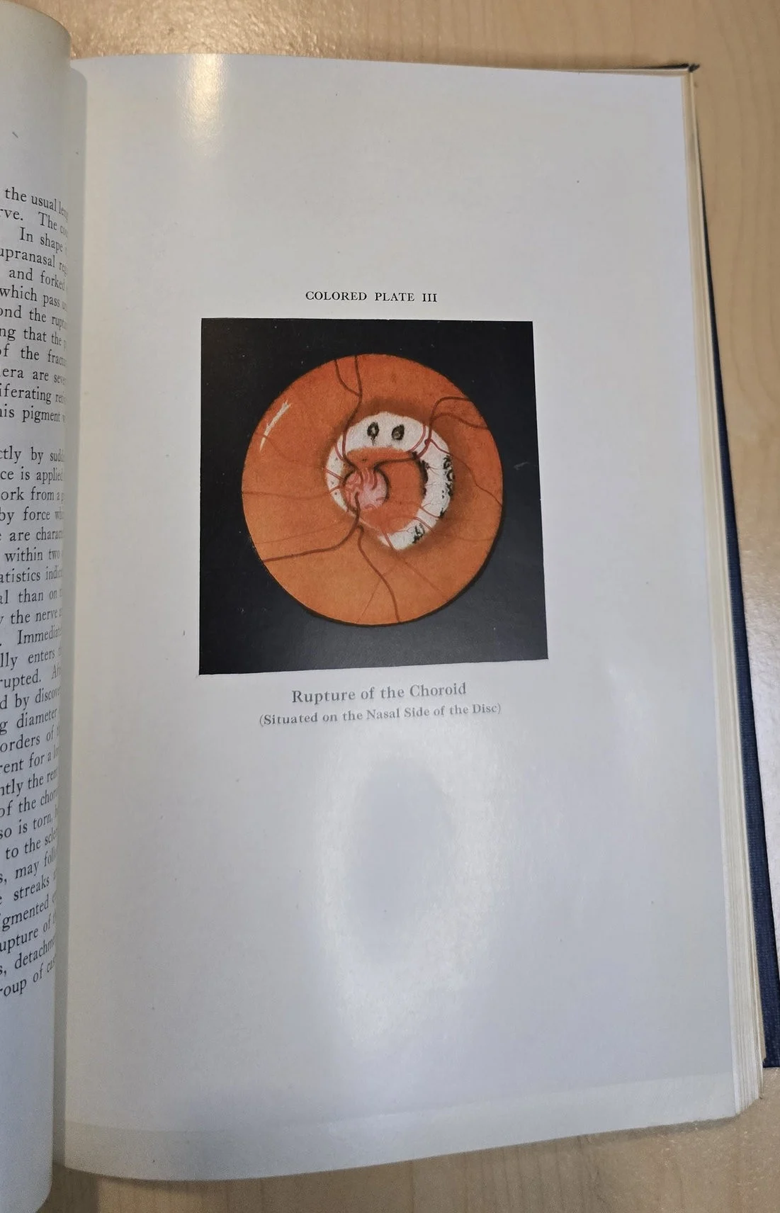
This extraordinary 1920 three-volume set is a pinnacle of early 20th-century ophthalmologic diagnostics. Authored by Dr. Edward L. Oatman, Diagnostics of the Fundus Oculi includes one bound text volume featuring 234 illustrations (including four stunning full-color plates), along with two matching diagnostic portfolios containing 79 stereograms and 8 diagnostic reference cards.
Each volume is a standalone teaching aid, designed to train physicians in the identification of retinal and optic disc abnormalities using direct examination and stereoscopic imaging—a precursor to modern fundus photography.
📚 Volumes in the Set
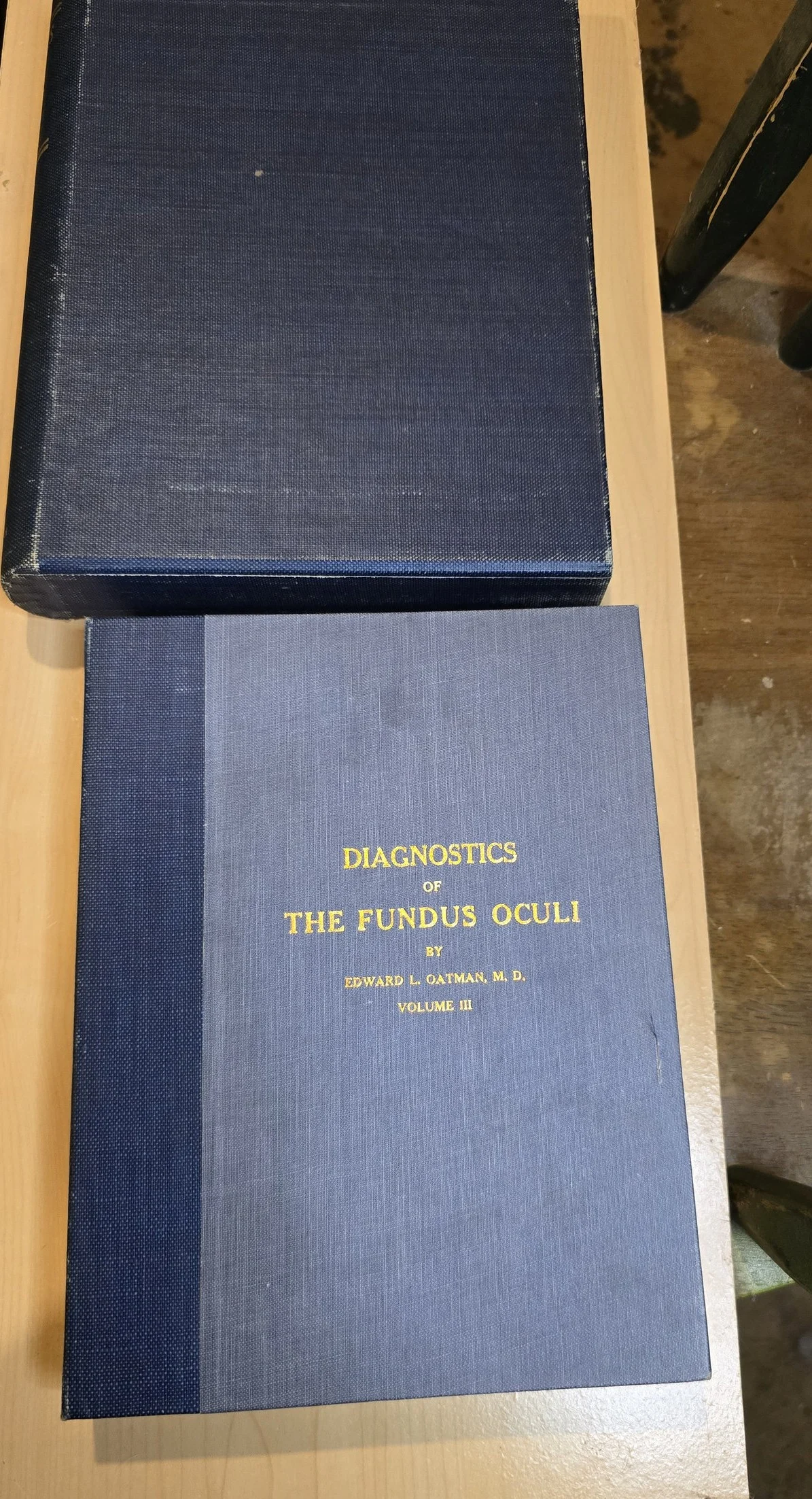
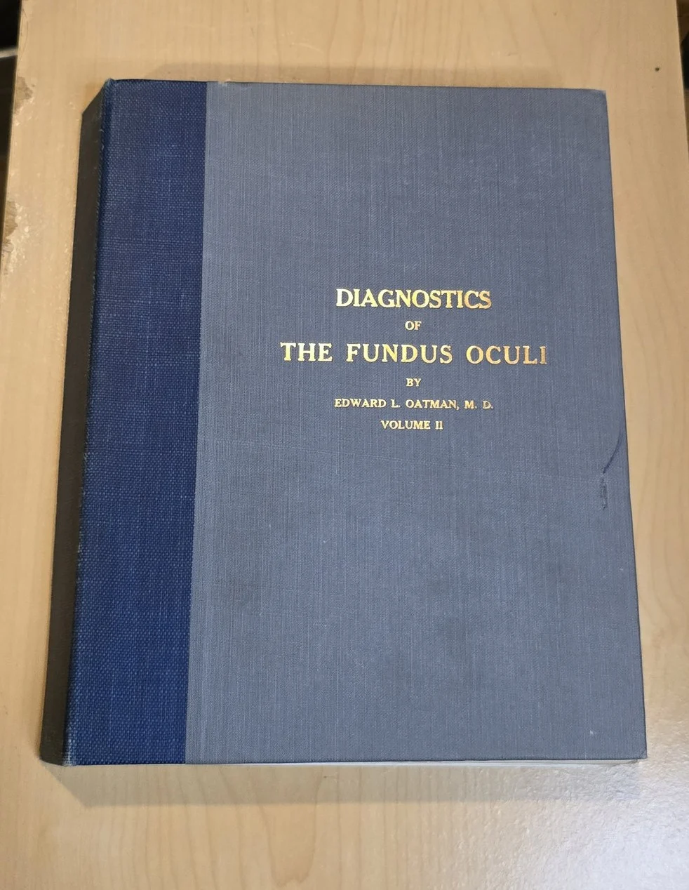
Volume I: Textbook
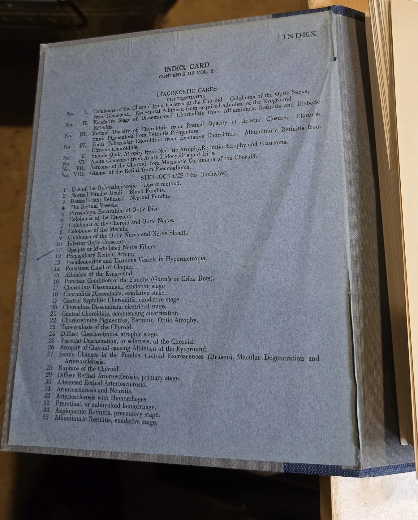
Hardcover, illustrated with extensive black-and-white diagnostic plates and four color illustrations.Volume II: Portfolio of Stereograms
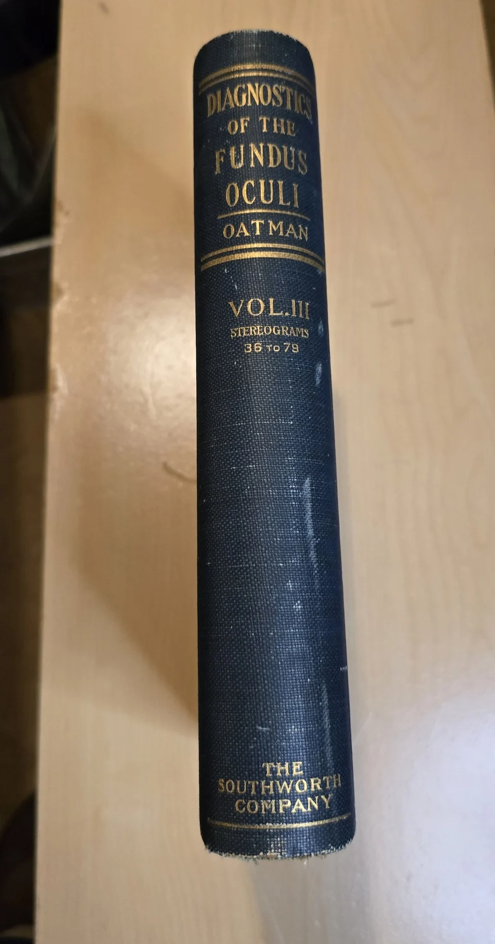
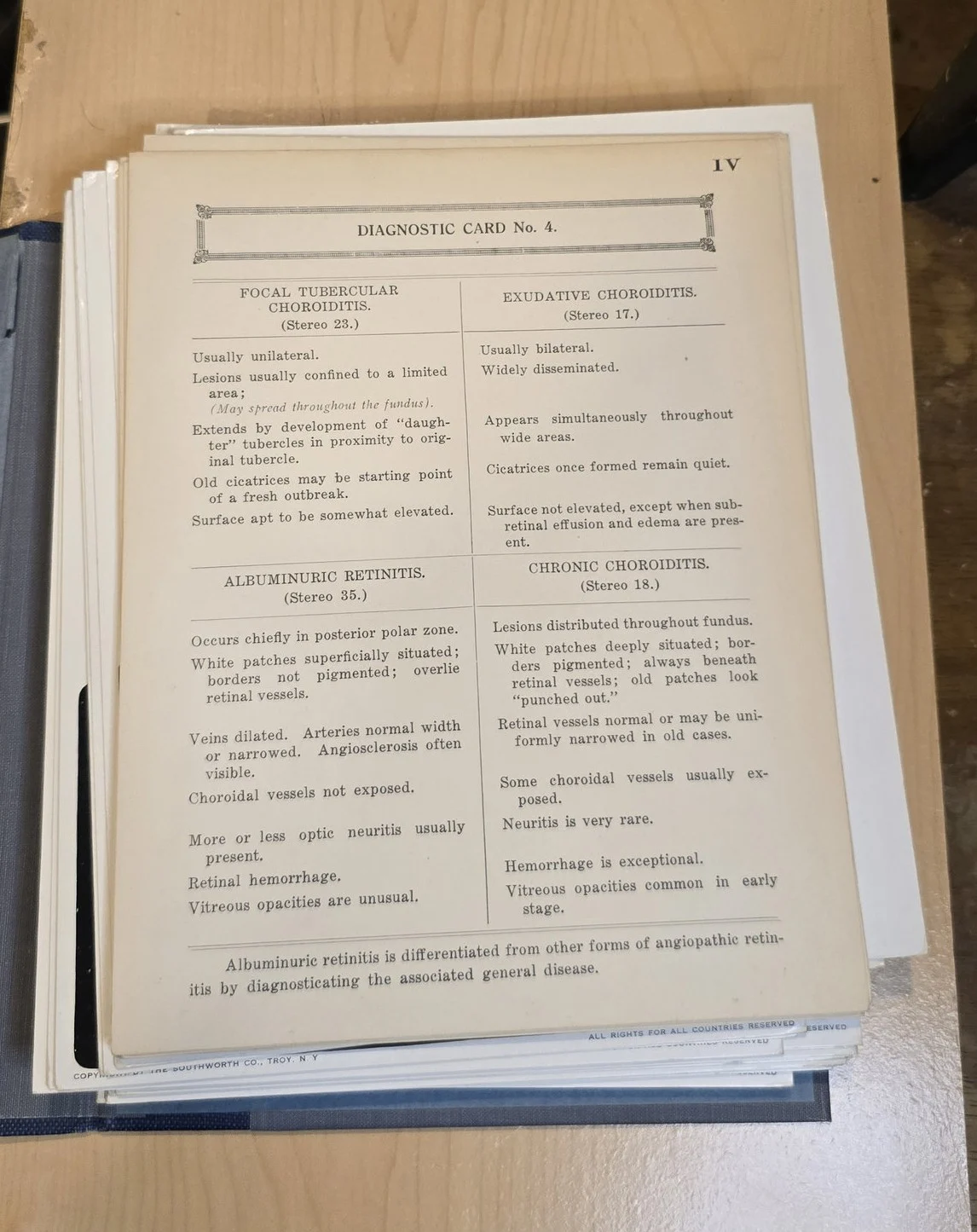
Loose stereoscopic cards with patient case details.Volume III: Portfolio of Diagnostic Cards
Reference cards for clinical identification of fundus pathologies.
(Both portfolios are housed in individual protective cases.)
Condition
Text volume: Excellent binding, clean pages, light edge wear
Portfolios: Complete, intact cards and stereograms
Cases: Mild rubbing and corner wear, labels intact
Images vivid, no major foxing, minor age toning
Gallery
Historical context
Published just after World War I, this set represents the golden age of observational medicine—before widespread retinal photography and optical coherence tomography (OCT). Physicians relied on direct ophthalmoscopy and stereoscopic interpretation to identify vascular diseases, optic atrophy, papilledema, and retinal hemorrhages. Oatman’s text and image portfolio helped standardize diagnostic interpretation during a crucial era in visual science.
Curious Facts, Ephemera, and Trivia
Fundus photography was still experimental in 1920; stereoscopic images were the gold standard for evaluating the optic nerve and retina.
The stereograms were meant to be used with a handheld stereoscope—bringing depth perception to pathology visualization.
This is one of the very few surviving complete sets with intact stereographic portfolios.
Excerpt
“Diagnosis must rely not upon the first glance at the fundus, but upon the trained appreciation of comparative subtlety—the true difference between vision and insight.”
Why it is in the Cabinet
This set is a cornerstone of early ophthalmic diagnostics, standing at the crossroads between anatomical observation and emerging technology. Its completeness—including fragile diagnostic cards and stereograms—makes it a true treasure in my Cabinet. The craftsmanship of the colored plates, paired with the clinical precision of the text, reflect a lost era of diagnostic elegance.
Support Dr. Bebout’s Cabinet of Medical Curiosities
If you enjoy the history, the oddities, and the effort, help keep this cabinet open. Every little bit helps preserve and share the strange wonders of medicine's past.
Buy Me a Ko-fi ☕ Buy Me a Coffee ☕ Tip via PayPal 💵