Title
Atlas and Epitome of Special Pathologic Histology
Author
Docent Dr. Hermann Dürck
Edited by Ludvig Hektoen, M.D.
Image
Description
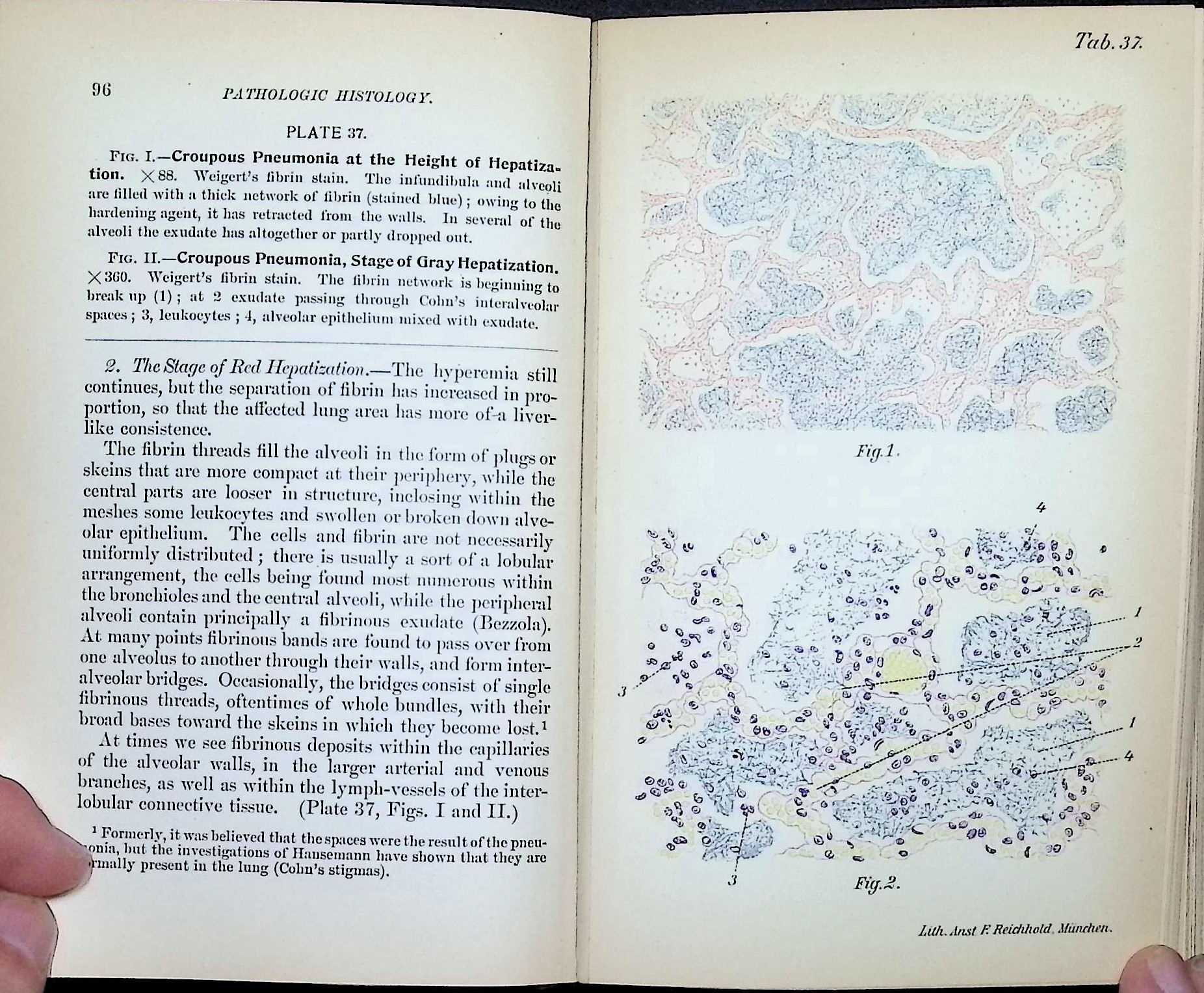
Published in 1900 by W. B. Saunders of Philadelphia, Atlas and Epitome of Special Pathologic Histology is an authorized English translation of Hermann Dürck’s German work, edited for American audiences by Ludvig Hektoen. The volume focuses on diseases of the circulatory system, respiratory organs, and gastrointestinal tract, presenting pathological changes through detailed text paired with finely executed color plates.
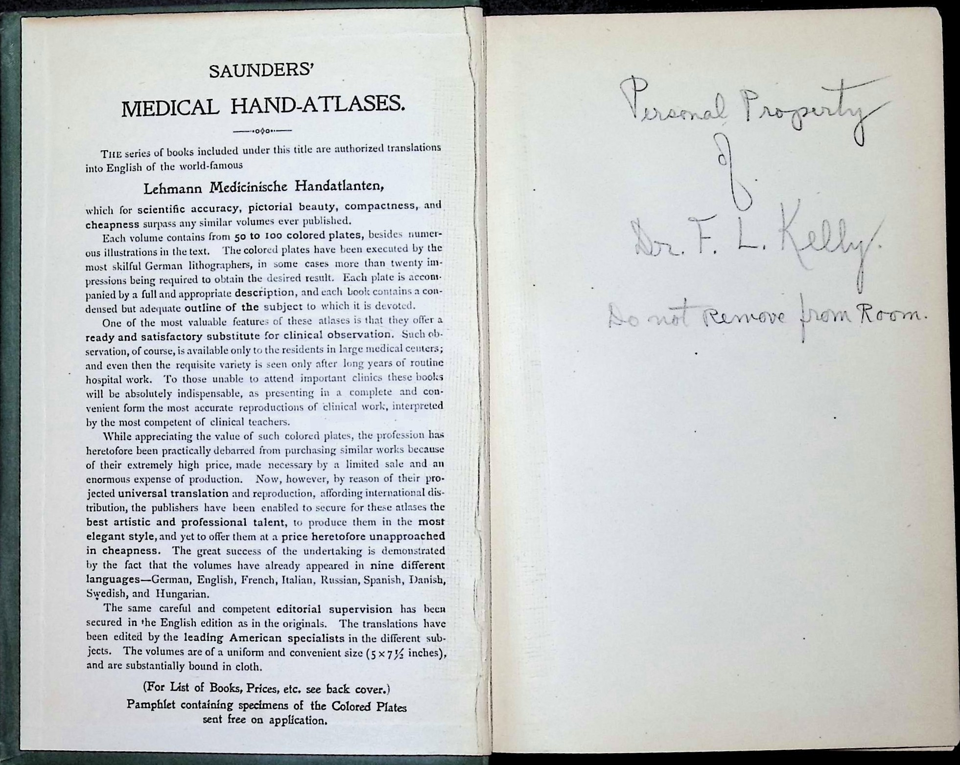
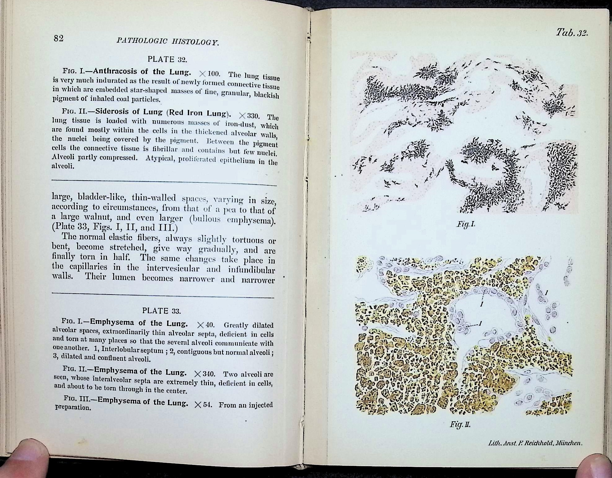
The book belongs to Saunders’ Medical Hand-Atlases series, which aimed to bring high-quality visual pathology references to medical students and physicians who lacked access to large teaching hospitals. Containing 62 colored plates, the atlas documents conditions such as amyloid degeneration, anthracosis, siderosis, emphysema, and other structural disease processes as they were understood at the turn of the 20th century.
Condition
Original green cloth binding with gilt spine lettering. Expected age-related wear present, including edge wear, surface scuffing, and small areas of cloth loss. Binding remains intact. Interior pages are clean and legible with light toning. Colored plates remain well preserved. Period ownership inscription present.
Gallery
Historical context
This volume reflects pathology at a pivotal moment: post-microscopy but pre-modern biochemistry and immunology. Disease is interpreted primarily through structural and morphological change, emphasizing gross and microscopic anatomy rather than molecular mechanisms. Atlases like this formed the backbone of pathology education before routine laboratory diagnostics and imaging.
Curious Facts, Ephemera, and Trivia
• Part of a larger multi-volume German atlas project later translated into several languages
• Saunders marketed these atlases as substitutes for direct clinical observation
• Many plates were produced using high-skill chromolithography
• Includes early visual descriptions of occupational lung disease
• Ownership note indicates use in a clinical or teaching environment
Excerpt
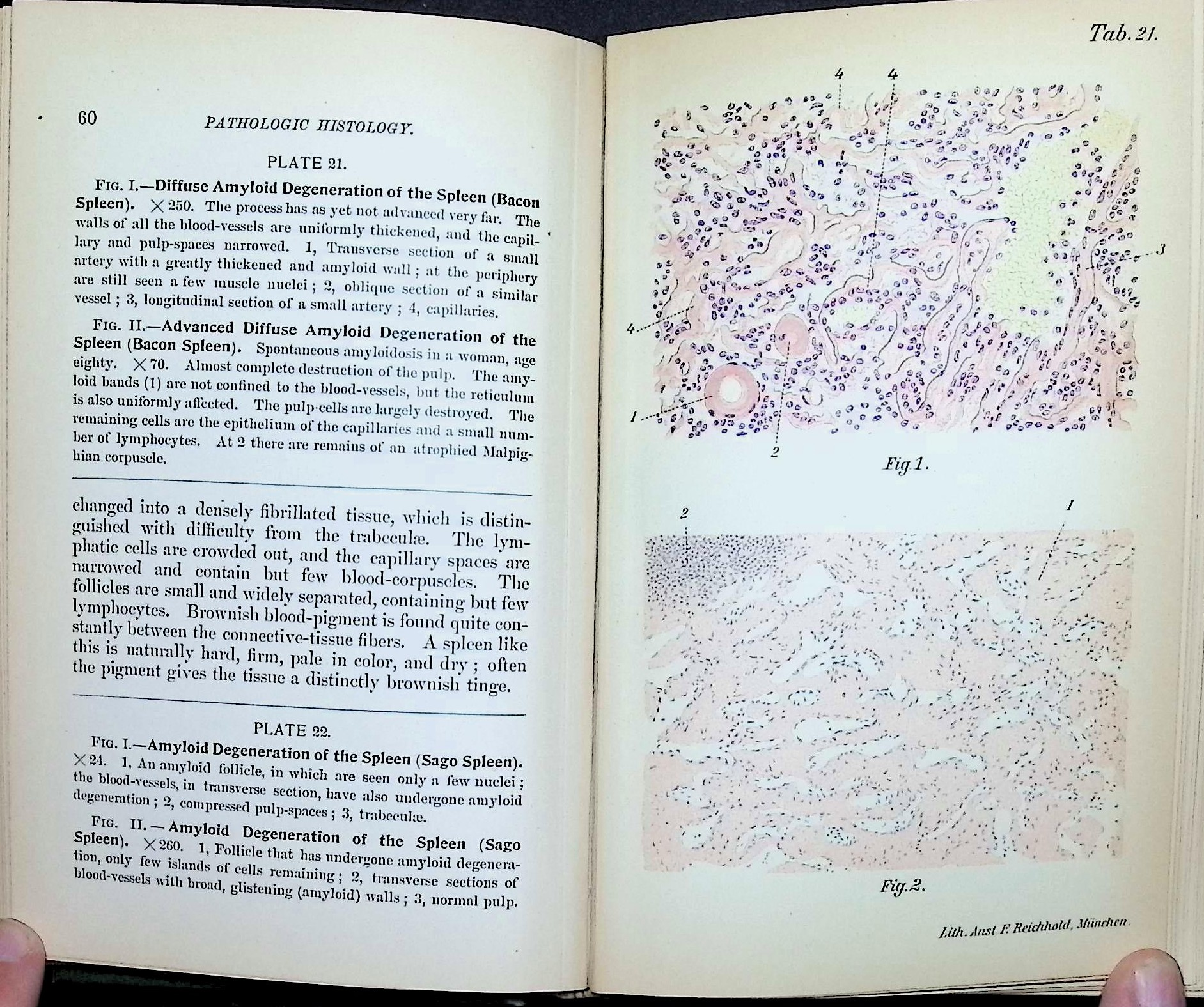
“Diffuse amyloid degeneration of the spleen… the walls of all the blood-vessels are uniformly thickened, and the capillary and pulp-spaces narrowed.”
Why it is in the Cabinet
This book captures pathology when diagnosis depended on the trained eye and the microscope alone. It represents the educational bridge between bedside observation and laboratory medicine, preserving how disease was seen before it could be measured. The clarity of its illustrations and the seriousness of its intent make it a foundational artifact in the history of medical education.
Support Dr. Bebout’s Cabinet of Medical Curiosities
If you enjoy the history, the oddities, and the effort, help keep this cabinet open. Every little bit helps preserve and share the strange wonders of medicine's past.
Buy Me a Ko-fi ☕ Buy Me a Coffee ☕ Tip via PayPal 💵